Fluorescence microscopy has been utterly transformative across countless biological investigative spheres by unveiling intricate tissue architectures, dynamic cell processes and molecular interactions otherwise wholly invisible under conventional brightfield imaging alone. This revolution stems from specialized fluorophores that tag proteins and cell elements which then emanate colorful luminescent glows when stimulated by specific excitation light wavelengths applied through the microscope optical path.

Researchers capitalize on this by labeling desired biological sample components using targeted fluorescent stains, antibodies or gene expressions to render key structures brilliantly visible or elucidate actively occurring cell physiology in action. Multi-color staining also enables characterizing complex interrelated particulate relationships. Thereby fluorescence methodologies unravel mysteries and phenomena fundamentally impossible to examine under standard microscopes, spawning once unimaginable research capabilities.
Understanding Fluorescence Microscopy Components and Principles
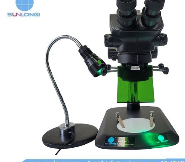
Standard fluorescence microscopes contain three primary elements working in concert to generate glowing luminescent emissions from target research specimens:
1. The Excitation Light Source
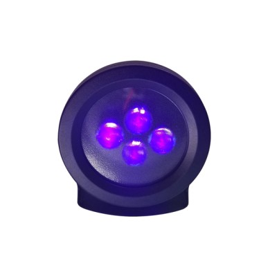
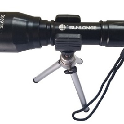
Sophisticated illumination towers like mercury lamps and LEDs produce high intensity spectra at defined wavelengths matching the peak excitation values of fluorescent molecules like DAPI or GFP commonly used in biological tissue labeling.
2. Specialized Emission Filters
Fluorescence barrier filters isolate and selectively permit passage of only the glowing wavelengths emitted from fluorophore probes once excited while blocking all other non-fluorescent light. This produces images absent background interference that would hinder contrast.
3. Microscope Objectives
Fluorescence microscope objectives maintain critical focus of both the excitation light and subsequent radiant emissions emanating from the fluorescently labeled sample, concentrating them through the optical lens systems into the observer’s eye or camera for clear bright imaging.
In conventional configurations, main objective ports connect directly to light sources. But installing a microscope fluorescence adapter between these two interfaces facilitates expanded functionality.
Core Benefits of Integrating Adapters
Fitting fluorescence adapters offers researchers two main advantages:
Multi-Modality Capacity Restoration
Adapters enable microscopes limited previously to just brightfield observation renewed fluorescence imaging capability that aids advanced cell and tissue investigations.
Operational Flexibility & Workflow Efficiency
Quick-change adapters mean easily alternating between routine and fluorescence microscopy modes on one body sans rerouting cables constantly between processes saving setup efforts. Adapters also allow implementing specialized techniques like FISH sequentially.
Additionally, both upright and inverted microscope configuration varieties interface with adapters making fluorescence more accessible for each orientation approach. This protects existing equipment asset investments if fluorescence becomes newly desired rather than necessitating entire integrated system replacements.
Vetting Key Adapter Specifications
With ever-growing excitement surrounding fluorescence imaging, numerous adapter variants now exist made by quality manufacturers. However, determining which aligns best to individual functionality needs means assessing available models across several parameters:
Spectral Range & Excitation Compatibility
Verify that the adapter offers excitation wavelength options compatible with your tower’s particular spectral output range and fluorophore labels used to sufficiently power quality emissions.
Microscope Mounting Orientations
Confirm the angled adapter design properly fits both vertically imposed paths on upright stands as well as right-angle transmissions through inverted scope specimen stages for reliable light relay with minimal leakage across space bound gaps.
Equipment Interfacing Standardization & Anti-Vibration
Check that adapter input/output threading and dimensions conform securely to existing equipment ports or component adapters without inadvertent instability that adds disruptive imaging artifacts over long-term routine use.
Required Imaging Channels
Determine necessary simultaneous channel imaging capacity specifications like 2-channel or 3+ channel based on planned fluorescence experimentation dyes and protocols ahead.
Imaging Clarity Assessment
Inspect sample specimen images through the fully integrated adapter at high 40X or 100X magnification for quality clarity, contrast uniformity, distortion absence or uneven field brightness that could indicate inadequate light optimization.
Construction Durability Factors
Seeking out metal construction generally proves wise for adapters over plastic versions more susceptible to accelerated photon-derived degradation that slowly impairs precision and quality output long-term under high-frequency usage demands.
Conclusion

Astutely adding adapters to infuse fluorescence capacity or optimize current systems unlocks transformational insights for tissue architecture and live cell dynamics analysis across countless research realms. But amidst the flourishing global adapter market, matching exact wavelength specifications, connectivity properties and optical path alignments requires diligent product comparisons against application-specific functionality needs. Consult closely with microscopy specialists at this important juncture to determine and integrate the optimal future-proof adapter ensuring maximal quantitative accuracy and streamlined investigative workflows amplifying unique discoveries for years ahead.
 CN
CN

